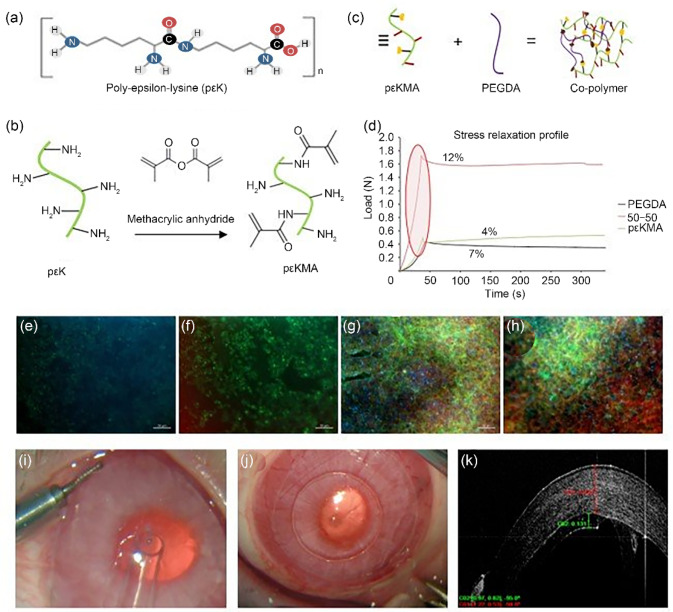
Fig. 6.
Fabrication of corneal hydrogels. a Poly-epsilon-lysine forms the basis of the hydrogels (pK). b pK is methacrylated by reacting m pK with methacrylic anhydride and triethylamine at pH 7 and 20 °C for 12 h to produce pKMA. Adapted from Ref. [108] (Copyright 2014, with permission from the authors, licensed under CC BY 4.0) and Ref. [109] (Copyright 2018, with permission from the authors, licensed under CC BY 3.0). c A co-polymer is formed from pK and poly(ethylene glycol) diacrylate (PEGDA). d The mechanical properties are improved for co-polymer hydrogels compared to pK and PEGDA alone. Cell attachment is affected by the formulation of the hydrogel as demonstrated using a human corneal endothelial cell line (HCEC12 cells). HCEC12 cells on e 100% pK hydrogel, f 90% pK/10% PEGDA, g 80% pK/20% PEGDA, and h 50% pK/50% PEGDA, after 7 d in culture (ZO-1 red, Phalloidin green, DAPI blue). The mechanical properties of the hydrogel are optimal for handling and delivery. i The hydrogel being delivered to the rabbit eye using an intraocular lens injector device and j after easy unfolding and centration in the anterior chamber. k The excellent positioning and attachment of the graft to the posterior rabbit cornea can be seen using optical coherence tomography (red indicating cornea and green graft). DAPI: 4',6-diamidino-2-phenylindole

