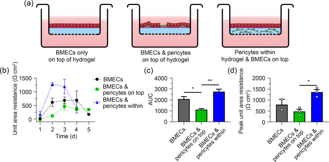
Fig. 7.
Pericytes embedded in hydrogel support the barrier function of brain microvascular endothelial cells (BMECs). OX1-19 induced pluripotent stem cells (iPSCs) were differentiated into BMECs and pericytes as described [112, 113]. PureCol hydrogel was layered on the surface of a Transwell insert with a coating of Matrigel on the upper surface. a BMECs were either cultured as a monoculture on top of the hydrogel, or mixed with pericytes on top of the hydrogel, or the pericytes were encapsulated within the hydrogel with the BMECs layered on top. Transendothelial electrical resistance (TEER) measurements were taken daily for five days and plotted as b unit area resistance (UAR) on respective days, c area under the curve (AUC) of b, and d peak UAR of b. TEER measurements were taken using an EVOM2 voltmeter and STX3 electrodes (World Precision Instruments, UK). Peak UAR is the highest recording of TEER of the whole measured time period. AUC and peak UAR were analysed using one-way analysis of variance (ANOVA) with Tukey’s multiple comparisons test. Data are shown as mean SEM. *P 0.05, **P 0.01. SEM: standard error of the mean

