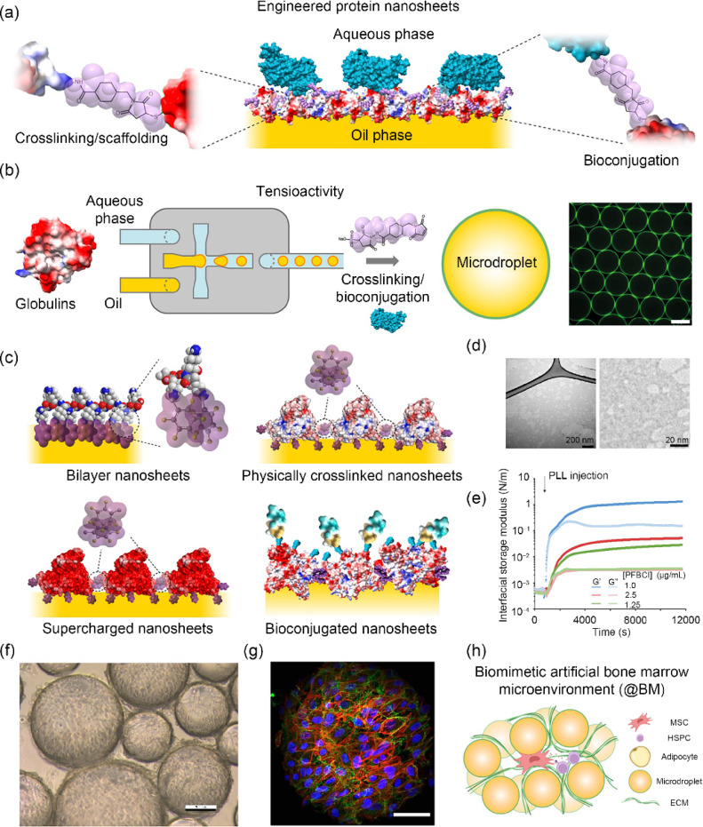
Fig. 8.
Biomanufacturing using microdroplet and protein nanosheet technologies. a Schematic representation of engineered protein nanosheets. Reproduced from Ref. [131], Copyright 2024, with permission from the authors, licensed under CC BY. b Protein nanosheet assembly can be orchestrated in microfluidic platforms to control the size of microdroplets. c Examples of protein nanosheet designs. Adapted from Ref. [124], Copyright 2023, with permission from the authors, licensed under CC BY 4.0. d Transmission electron microscopy images of protein nanosheets (albumin-based). e Changes in interfacial shear mechanics taking place upon assembly of poly(L-lysine) nanosheets at a liquid–liquid interface. Reproduced from Ref. [122], Copyright 2022, with permission from the authors, licensed under CC BY 4.0. f Brightfield microscopy image of HEK293 cells growing on a bioemulsion. g Colony of mesenchymal stem cells growing on a microdroplet (blue, nuclei; red, F-actin; green, vinculin). Reproduced from Ref. [123], Copyright 2023, with permission from the authors, licensed under CC BY 4.0. h Schematic representation of a microdroplet-based bone marrow microenvironment. Reproduced from Ref. [133], Copyright 2023, with permission from the authors, licensed under CC BY 4.0

