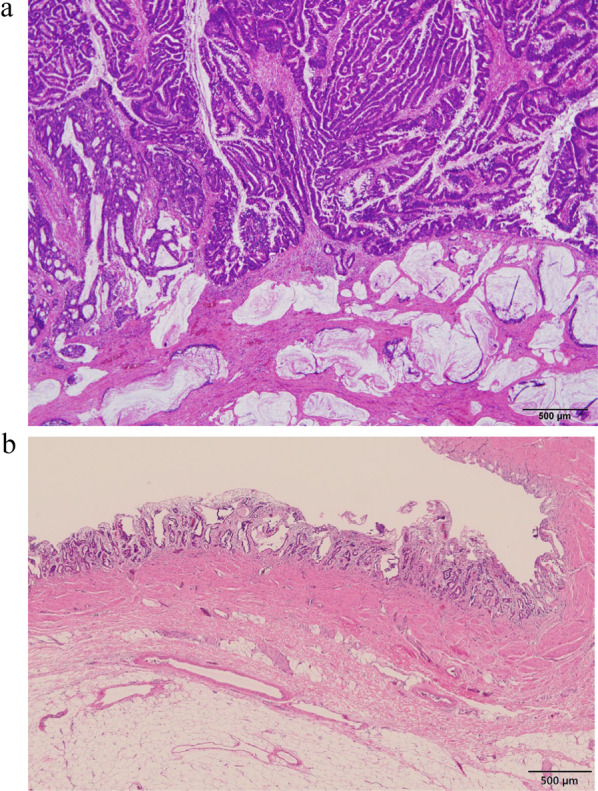
Fig. 3.

Pathological findings. Histopathologically, adenocarcinoma was located mainly in the right hepatic and common bile ducts (innumerable tumors, maximum diameter: 5.6 cm, papillary-infiltrating type, papillary adenocarcinoma > moderately differentiated tubular adenocarcinoma > mucinous adenocarcinoma). No vascular invasion was observed. Lymphatic metastasis was observed in a lymph node on the posterior surface of the pancreatic head (pathologically minimal lymphatic invasion). a The tumor invaded the bile duct wall and the liver parenchyma in some areas. b Biliary intraepithelial neoplasia was also observed in the left hepatic duct. Pathological diagnosis: pT2b, pN1, M0, Stage IIIc (UICC, 8th edition)
