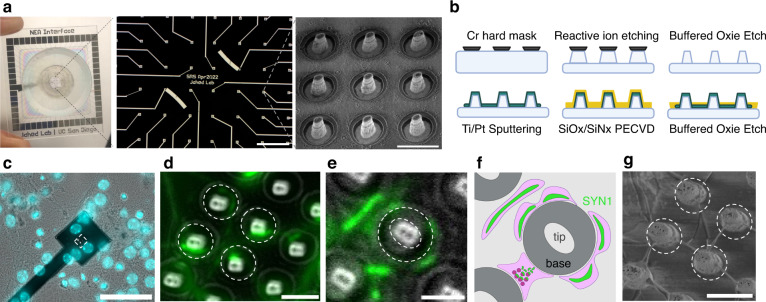
Fig. 1. NEA fabrication and nanopillar interactions with neuron-like cells.
a Schematic showing 4 cm × 4 cm NEA containing 60 total electrodes, each containing 9 nanopillars ~3 μm tall and 600 nm wide at the tip (scale bars: 500 μm, 5 μm). b Schematic summarizing NEA fabrication, consisting of two-step wet and dry etching and standard photolithography to pattern electrodes. c Representative fluorescence image with NucBlue staining of a single nanoelectrode showing sparse interactions between primary neuron somas and nanopillars (scale bar: 50 μm). d Merged fluorescence image, with the white dashed circles indicating 4 different nanopillars (scale bar: 3 μm), showing synapsin-1 punctae curving around separate nanopillars. e Another merged fluorescence image showing synapsin-1 punctae curving around a single nanopillar (scale bar: 2 μm). The white dashed circles indicate the tip and base of the nanopillar. This same pillar is depicted schematically in (f), demonstrating putative microscale curvature achieved by synapsin-1 distribution across the presynaptic region of the neurite. Created with BioRender.com. g Zoomed in SEM images of ROI shown in (d) with white dashed circles corresponding to the same four nanopillars (scale bars: 3 μm)

