
FIGURE 5.
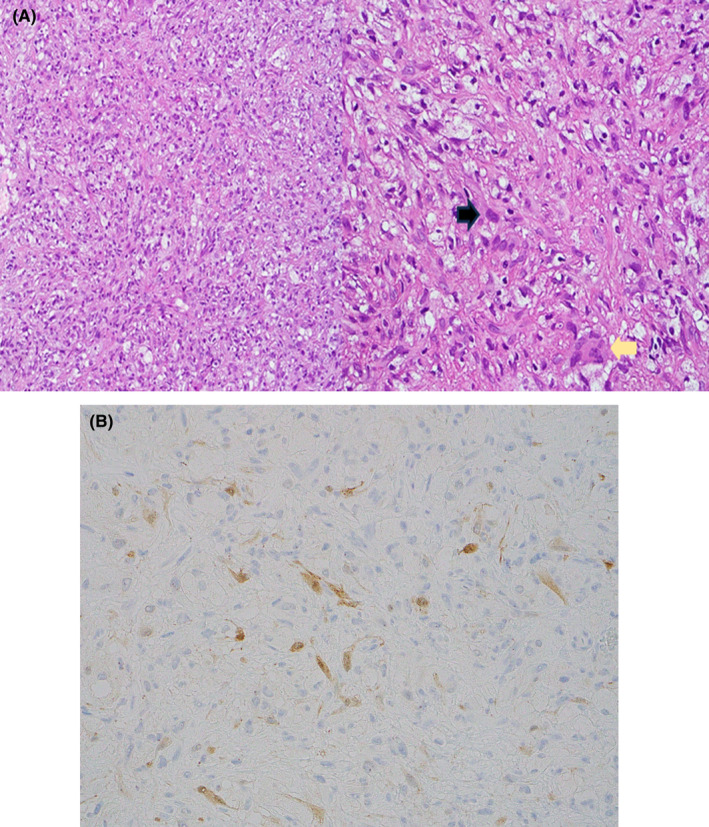
(A) Light microscopic examination reveals a tumor characterized by a proliferation of xanthomatous histiocytes with smaller, moderately atypical epithelioid cells with eosinophilic cytoplasm (black arrow). Additionally, a few osteoclast giant cells were noted (yellow arrow) (H&E stain, magnification × 100, × 400). (B) Isolated epithelioid cells were highlighted by keratin immunohistochemistry (H&E stain, × 400).