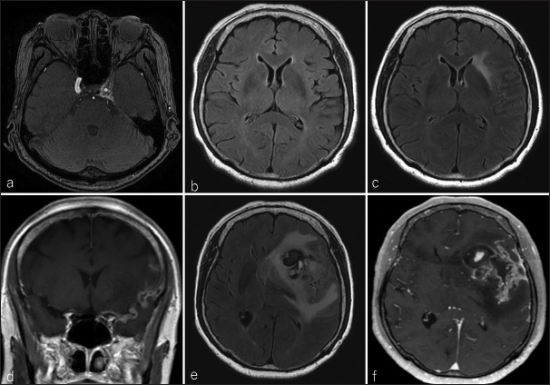
Figure 1:

(a) Magnetic resonance angiography in the axial view revealed a left internal carotid cavernous sinus fistula (CCF) 13 years before the presentation; (b) imaging 1 year before presentation showed no occurrence of CCF or abnormal signal in the left subventricular zone (SVZ). (c) At presentation, the SVZ was highly intense on fluid-attenuated inversion recovery (FLAIR), and (d) extensive leptomeninges from the basiler cistern to the left Sylvian fissure could be observed on contrast-enhanced T1 weighted imaging (CE-T1WI). After 2 months, the tumor was enlarted and bled on (e) FLAIR and (f) CE-T1WI images.
