Figure 2:
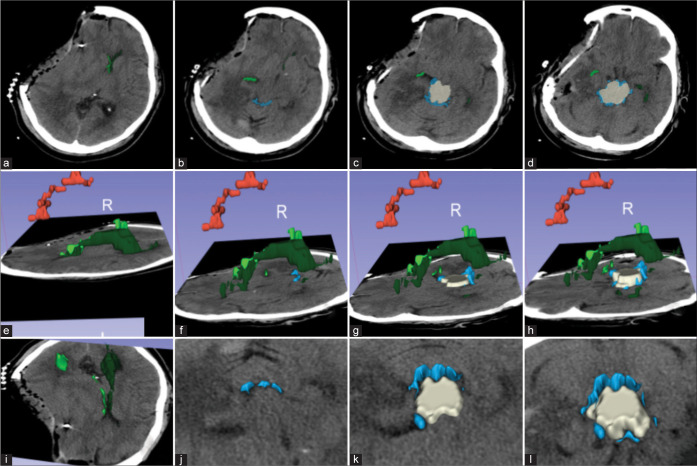
Assessment of postoperative changes on the same patient as Figure 2. (a) Axial images showing relief of midline shift over the right (light green) and left (dark green) lateral ventricles and a new right cranial defect after a decompressive hemicraniectomy, and (b-d) improvement of the visibility of the perimesencephalic cisterns (blue sky) and midbrain (beige); (e-h) 3D reconstruction with a left lateral view showing residual subdural hematoma and improvement in ventricular diameter; (i-l) A frontosuperior view of the axial images showing improvement over the midbrain compression and perimesencephalic cisterns. Red: Subdural hematoma; beige: midbrain; light green: right lateral ventricles; dark green: left lateral ventricles; blue: perimesencephalic cisterns.

