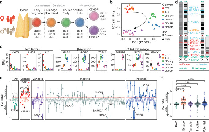
Fig. 1. Sex-biased escape gene expression in human thymocytes.
a Thymocyte development and markers used for cell sorting. b Principal component analysis (PCA) of normalized RNA-seq counts from male (triangle) and female (circle) developing thymocytes. c Expression dynamics as transcript per million (TPM) of genes associated with key transitions during thymocyte development. Boxplot representing median (central line), first and third quartiles (Q1 and Q3, respectively) (box edges) and 1.5*inter quartile range (IQR) from Q1 and Q3 (whiskers) from seven biological replicates (3 males and 4 females) are shown. d Schematic representation of selected genes escaping and inactivated by X-chromosome inactivation on the active (Xa) and inactive (Xi) X-chromosome. e Sex-biased expression of genes across the X-chromosome as log2 fold change (FC) of expression in female over male thymocytes. Horizontal red lines indicate median (dotted line) ± IQR of inactive genes (filled lines). FC (log2) values have been capped to 1 or −1 if above or below ±1, respectively. FC (log2) (dots) and standard error (vertical lines) of seven thymocyte populations from females (n = 4) and males (n = 3) shown. f Sex-biased gene expression across the X-chromosome as log2 FC of expression in female over male thymocytes by known XCI status. P-values from Benjamini-Hochberg (BH) corrected two-tailed T-test. Boxplot representing median (central line), Q1 and Q3 (box edges) and 1.5*IQR from Q1 and Q3 (whiskers) across all genes. Dots representing mean of six thymocyte populations from seven biological replicates (3 males and 4 females). e, f XCI status defined based on previous assessment10 with potential XCI escape genes (blue) previously not investigated or classified as unknown. a, b, c ETP, early T cell progenitors; T-C, T cell committed thymocytes; DPearly, early double positive thymocytes; DPlate, late double positive thymocytes; CD4SP, CD4 single positive thymocytes; CD8SP, CD8 single positive thymocytes. d, e, f PAR, pseudoautosomal region. Source data are provided as a Source Data file.

