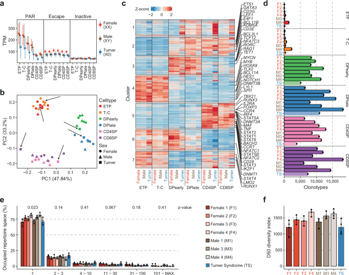
Fig. 8. The inactive X-chromosome does not contribute to T cell development in humans.
a Expression as transcript per million (TPM) of genes in PAR (pseudoautosomal region), escape and inactive genes for each thymocytes subpopulation in XX females (orange, 4 biological replicates), XY males (brown, 3 biological replicates) and X0 Turner syndrome patient (blue, 1 biological replicate). Points indicate mean and lines standard error. b Principal component analysis (PCA) of normalized RNA-seq counts from developing thymocytes from XX and XY karyotype females (circle) and males (triangle) as well as a Turner syndrome patient (TS) (square). TS samples are highlighted with black arrows. c Average Z-score per karyotype and cell type for each cluster of dynamic gene expression throughout thymocyte development identified in Supplementary Fig. 2c. d Clonotype count for thymocyte subpopulations in females (F1-F4), males (M1, M3, M4) and a Turner syndrome patient (TS). e Occupied repertoire space of clonotypes with 1, 2-3, 4-10, 11-30, 31-100 and 101-MAX number of clones in female, male and TS samples. P-values (Kruskal-Wallis test with adjustment for multiple testing by Holm) are indicated at the top. f Diversity score in DPearly, DPlate, CD4SP and CD8SP cells of F1, F2, F3, F4, M1, M3, M4 and TS. e, f Bars indicate mean and error bars 95% CI of four thymocyte subpopulations (DPearly, DPlate, CD4SP, CD8SP) for each individual. a, b, c, d, ETP, early T cell progenitors; T-C, T cell committed thymocytes; DPearly, early double positive thymocytes; DPlate, late double positive thymocytes; CD4SP, CD4 single positive thymocytes; CD8SP, CD8 single positive thymocytes. Source data are provided as a Source Data file.

