Figure 4:

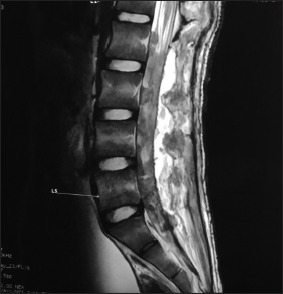
Postoperative contrast image with visible nerve roots at L1–L2 levels with comparatively lesser enhancement as compared to the preoperative state, and laminectomy defects visible. Arrow: L5 Vertebral level.

Postoperative contrast image with visible nerve roots at L1–L2 levels with comparatively lesser enhancement as compared to the preoperative state, and laminectomy defects visible. Arrow: L5 Vertebral level.