. 2024 Dec 5. Online ahead of print. doi: 10.1039/d4tb01334d

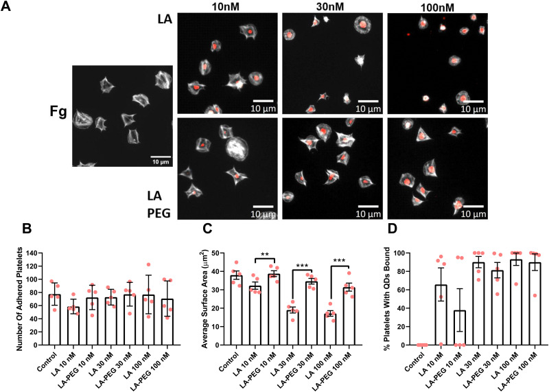
Fig. 6. Platelet spreading is maintained in the presence of PEGylated LA QDs. Washed platelets at 2 × 10⁷ mL−1 were spread on 100 μg mL−1 fibrinogen (Fg) in the absence, or presence of either QD-LA or QD-LA-PEG at 10 nM, 30 nM or 100 nM for 25 min before permeabilizing, fixing and staining with FITC-Phalloidin. Coverslips were mounted and visualized by fluorescence microscopy using an NA 1.4 oil x63 objective. (A) Representative images with scale bars showing 10 μm. Graphs show (B) number of adhered platelets, (C) average surface area of platelets (μm2), and (D) percentage of platelets with QDs bound. Data is shown as mean ± SD. n = 5. Statistical significance was calculated using a two-way ANOVA followed by Sidak's multiple comparison post hoc test. *p ≤ 0.05, **p ≤ 0.01, ***p ≤ 0.001.

Fig. 6