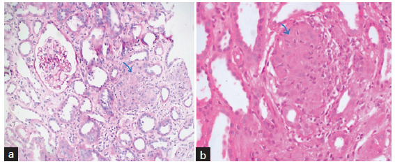
Figure 2:

(a) Section from renal cortex shows circumscribed non-necrotizing granuloma comprising epithelioid histiocytes and lymphocytes (blue arrow). PAS stain ×200 original magnification. (b) Section from the renal cortex shows circumscribed non-necrotizing granuloma composed of epithelioid histiocytes and lymphocytes (blue arrow) in another patient. H&E stain ×400 original magnification.
