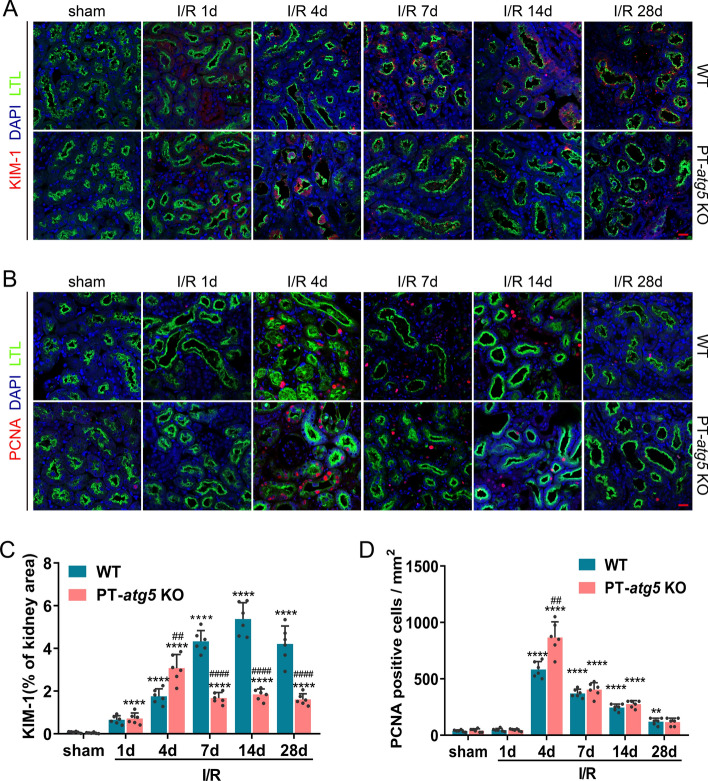
Fig. 4.
Dynamic changes of KIM-1 and PCNA in Atg5-deficient proximal renal tubules after I/R injury. WT and PT-atg5 KO mice kidneys were harvested at 1, 4, 7, 14, and 28 days after sham surgery or bilateral ischemia/reperfusion, as described in Fig. 1, to observe the dynamic course of kidney injury and repair. A Immunofluorescence staining of KIM-1 (red) in the kidney sections. B Representative PCNA (red) immunofluorescence staining of kidney sections. Kidney tissue sections were stained with DAPI to visualize cell nuclei (blue) and LTL to visualize proximal tubules (green). Scale bars, 20 μm. C Quantitative analysis of the KIM-1 positive stained areas. D Quantification of PCNA-positive cells per mm2. The dates are shown as mean ± SD (n = 6). * Indicates significant difference from the sham group; # indicates a significant difference from the relevant wild-type group. # or *P < 0.05, ## or **P < 0.01, ### or ***P < 0.001, #### or ****P < 0.0001

