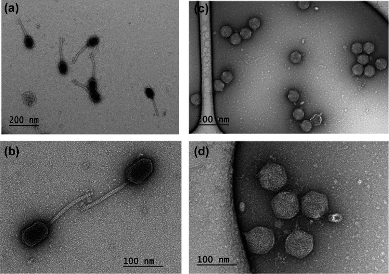
Fig 1.
Transmission electron micrographs of ZC01 and ZC03 purified phage particles, negatively stained with uranyl acetate. Different areas on a grid show intact ZC01 (a and b) and ZC03 (c and d) phage particles with siphovirus (ZC01) and podovirus (ZC03) morphotypes as previously described (46).

