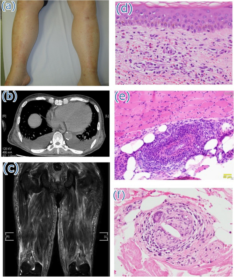
Fig. 1.

a Physical examination findings. Purpura on the lower legs is observed. b Contrast-enhanced computed tomography of the chest and abdomen reveals calcification of the descending aorta. c Magnetic resonance imaging of the thighs reveals diffuse intramuscular hyperintensities on T2-weighted images and short tau inversion recovery sequences in the hamstrings and quadriceps femoris. d Histological findings of the purpura on the lower leg demonstrate perivascular inflammatory cell infiltration with hemorrhage and nuclear dust. e Histological examination of the left vastus lateralis muscle reveals two vessels surrounded by lymphocytes and macrophages, with no evidence of fibrinoid necrosis. f Random skin biopsy shows the presence of crystal clefts within small blood vessels in the superficial dermal layer
