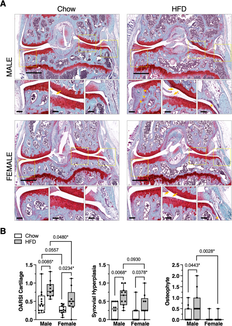
Fig. 5.
Male and female mice develop greater knee OA pathology with HFD versus chow. A Representative histological images of mid-coronal knee sections stained with Safranin-O, fast green, and hematoxylin from male and female mice fed Chow or HFD. Large panel scale bar = 500 μm. Dashed boxes in large panel indicate locations selected for small panel images representing, from left to right, the medial joint margin, lateral femoral-tibial cartilage loading region, and synovial lining inferior to the lateral meniscus. In small panels, arrowheads indicate osteophyte development (male HFD), arrows indicate mild cartilage damage (male Chow, male HFD, female HFD), and asterisks indicate synovial thickening (male HFD, female HFD). Small panel scale bars = 100 μm. B Semi-quantitative histological grading of cartilage, synovium, and osteophyte pathology. All scoring was performed by 2 experienced graders in a blinded manner. OARSI cartilage score range = 0–6; Synovial hyperplasia score range = 0 (absent) or 1 (present); Osteophyte score range = 0–3. Data points represent average values for individual animals. Boxes represent the 25th to 75th percentiles, horizontal line indicates the median, and whiskers demonstrate maximum and minimum values. Data were analyzed by Kruskal-Wallis test: OARSI cartilage (p = 0.0002), Synovial hyperplasia (p = 0.0008), Osteophyte (p = 0.0035). Post-hoc paired comparisons based on two-stage linear step-up procedure of Benjamini, Krieger and Yekutieli to control for 10% false discovery rate, q (p < 0.10 shown; *q < 0.10). Sample sizes for each analysis provided in Supplemental Table S3

