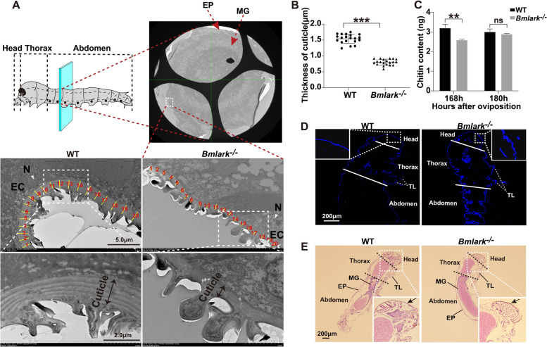
Fig. 2.
Structural differences in the cuticle of WT and Bmlark−/− embryos at the head pigmentation stage. A TEM images of the cross sections of WT and Bmlark−/− embryos at the head pigmentation stage (168 h post oviposition). The upper part of the figure shows the crosscut position of the embryo, along with a TEM image of the entire associated transverse section (original images are presented in Supplementary Figure S2A). The cyan plane represents the cut position, which is consistent in both WT and Bmlark−/− embryos, and located roughly in the second segment of the abdomen. EP: epidermis. MG: midgut. EC: epidermal cell. N: nucleus. The yellow lines, which are perpendicular to the tangent lines along the cuticle, represent the thickness of the cuticle at 20 randomly selected regions. B Comparison of the thickness of the WT and Bmlark−/− cuticle in the epidermis of the abdomen. The length of yellow lines at 20 different regions per individual in Figure (A) were calculated using Nano Measurer 1.2 software, and represent the thickness of the cuticles in the epidermis of WT and Bmlark−/− embryos. Student’s t test was used to evaluate statistical significance. Results are presented as the mean ± SE. ***p ≤ 0.001. C Chitin content of WT and Bmlark−/− embryos 168 and 180 h post oviposition. The ordinate represents the mass of chitin per gram of embryonic tissues. Student’s t test was used to evaluate statistical significance. Results are presented as the mean ± SE of three biological replicates. **p ≤ 0.01; ns: no significance, p > 0.05. D Chitin staining of the sagittal sections of the WT and Bmlark−/− mutant embryos at 180 h post oviposition. The white lines divide different parts of the body. Chitin was stained blue with fluorescent brightener 28. TL: thoracic leg. (E) HE stained images of the sagittal sections of the WT and Bmlark−/− embryos at 180 h post oviposition. The arrows indicate the head cuticle. The black dotted linesdivided different parts of the body. MG: midgut. EP: epidermis. TL: thoracic leg. Enlarged original images of the head of WT and Bmlark−/− embryos are presented in Supplementary Figure S2B

