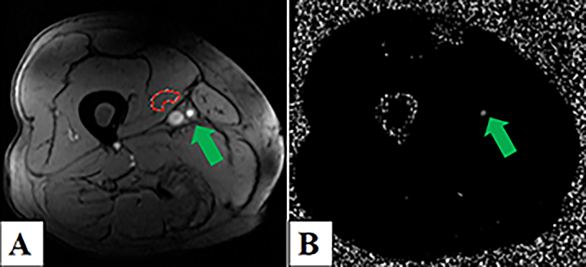
Figure 1.

Two-dimensional-phase-contrast magnetic resonance imaging (2D-PC-MRI) of the right distal superficial femoral artery (SFA) in a non-diabetic peripheral artery disease (PAD) patient. Panel A) Magnitude image depicting the background correction region of interest in the vastus medialis (red contour) and the SFA (green arrow). Panel B) Corresponding phase-contrast image showing the SFA (green arrow).
