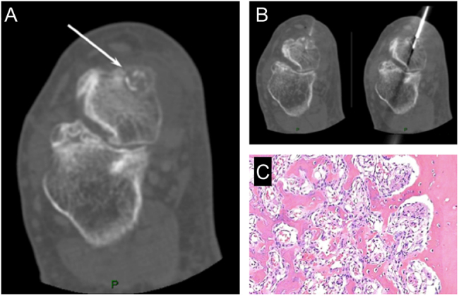
Figure 5.

Osteoid osteoma of the talus in a 45-year-old male. (A) CT scan clearly shows the nidus (white arrow); (B) CT scan during different phases of the thermoablation. (C) Histology shows trabeculae of woven bone of variable thickness, fibrovascular stroma, a well-defined border of the lesion and a dense sclerotic reactive border.

 This work is licensed under a