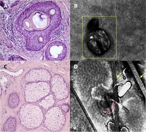
Figure 2.

RCM evaluation and related histopathological section of alopecia areata incognita: infundibular ostia empty or full of sebum and keratin (A), visualized at RCM as an infundibulum fulfilled with bright material which corresponds to keratin (yellow square - dermis) (B), miniaturized anagen-vellus hair follicles (“nanogen”) (C), seen in RCM as miniaturized follicles, represented by a reduction of the caliber of the hair shaft (pink arrows) in comparison to normal hair (yellow arrows - corneal layer) (D) (H&E, ×8).
