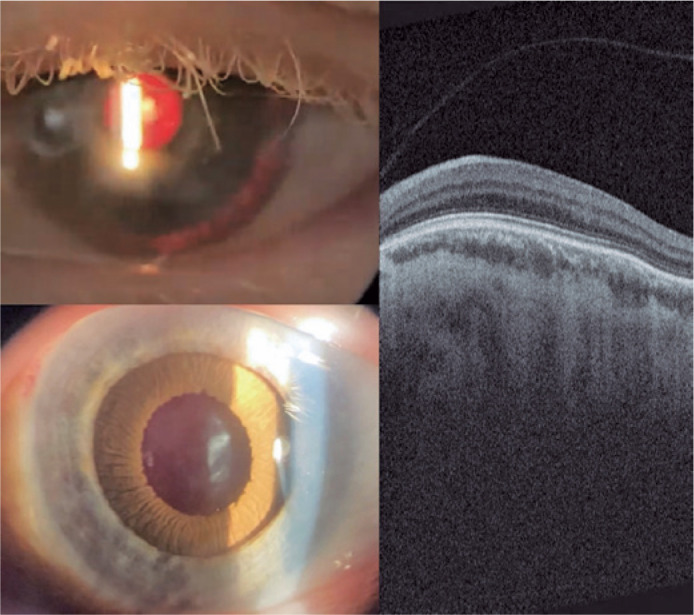
Figure 4.

Left-eye slit lamp photograph 30 days after surgery, showing prosthetic iris on ciliary sulcus through retro illumination and after pharmacologic mydriasis. Retinal OCT showing absence of foveal pit and persistence of retinal inner layers through expected area of fovea (macular hypoplasia).
