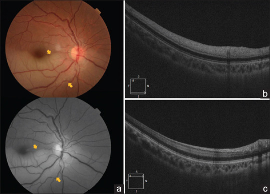
Figure 1.

(a) Color and red-free fundus photo showing inferotemporal pale retina due to BRAO in the right eye (yellow arrows). (b) Optical coherence tomography (OCT) scan of the right eye showing hyperreflective band in the inner plexiform and inner nuclear layer and thickening of the retinal layer. (c) OCT scan of the corresponding superior arcuate area shows normal thickness and reflectivity of inner retinal layers
