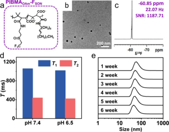
Figure 4.

(a) Chemical structures of PIBMAOAm-FSON. TEM image (b) and 19F NMR spectrum (c) of PIBMAOAm-FSON-IR780 NPs. (d) Comparison of T1 and T2 of PIBMAOAm-FSON-IR780 NPs at different pH values. (e) Evolution of particle size along with incubation time. Inset photographs were PIBMAOAm-FSON-IR780 NPs powder and PIBMAOAm-FSON-IR780 NPs redispersed in PBS.
