. 2024 Dec 5;13:RP100569. doi: 10.7554/eLife.100569

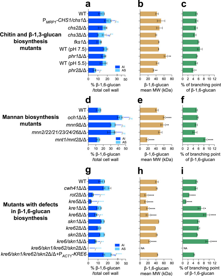
Figure 3. Comparative analysis of β-1,6-glucan content and structure produced by cell wall mutants.

(a, d, g) Percentages of cell wall β-1,6-glucans (alkali-insoluble [AI] and alkali-soluble [AS] fractions) on total cell wall. (b, e, h) β-1,6-Glucans mean molecular weight (MW). (c, f, i) Branching rate of β-1,6-glucans. Cells were grown in liquid synthetic dextrose (SD) medium at 30°C. Means and standard deviations from three independent replicate experiments are shown. All data were compared to the control conditions and were analyzed by one-way ANOVA with Dunnett’s multiple comparisons test: *p<0.05; **p<0.01; ***p<0.001; ****p<0.0001; ns, nonsignificant; NA, nonapplicable.

Figure 3—source data 1. Raw data for panels a to i.

Figure 3.

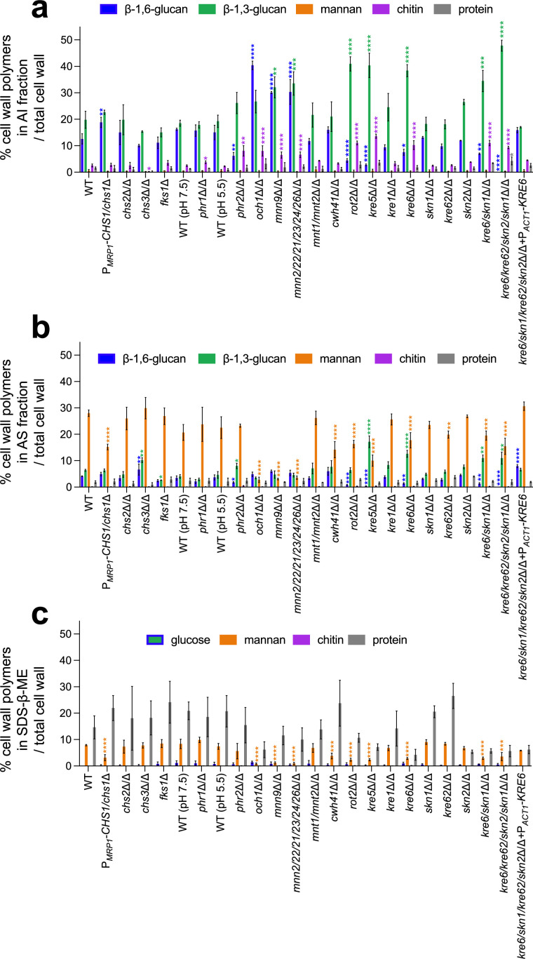
Figure 3—figure supplement 1. Cell wall composition of C. albicans mutants.

Figure 3—figure supplement 1.

Results are represented as the percentage of each polymer on total cell wall in alkali-insoluble (AI) fraction (a), alkali-soluble (AS) fraction (b), and sodium-dodecyl-sulfate-β-mercaptoethanol (SDS-β-ME) fraction (c). Means and standard deviations were obtained from three independent replicate experiments. All data were compared to the parental strain SC5314 and were analyzed using one-way ANOVA with Dunnett’s multiple comparisons test: *p<0.05; **p<0.01; ***p<0.001; ****p<0.0001.
Figure 3—figure supplement 1—source data 1. Raw data for panels a, b and c.
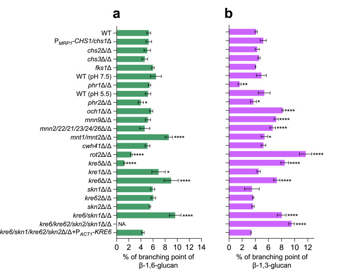
Figure 3—figure supplement 2. Branching rates of β-1,6-glucans and β-1,3-glucans produced by different cell wall mutants of C. albicans.

Figure 3—figure supplement 2.

(a) Branching rate of β-1,6-glucans from alkali-insoluble (AI) fraction. The branching rate was estimated by high-performance anion exchange chromatography (HPAEC) after digestion by an endo-β-1,6-glucanase. (b) Branching rate of β-1,3-glucans from AI fraction. The branching rate was estimated by HPAEC after digestion by an endo-β-1,3-glucanase. Means and standard deviations from three independent replicate experiments are shown. All data were compared to the control conditions and were analyzed using one-way ANOVA with Dunnett’s multiple comparisons test: *p<0.05; **p<0.01; ***p<0.001; ****p<0.0001; NA, nonapplicable.
Figure 3—figure supplement 2—source data 1. Raw data for panels a and b.
Figure 3—figure supplement 3. Absence of β-1,6-glucans in the cell wall of the quadruple kre6/kre62/skn2/skn1Δ/Δ mutant.
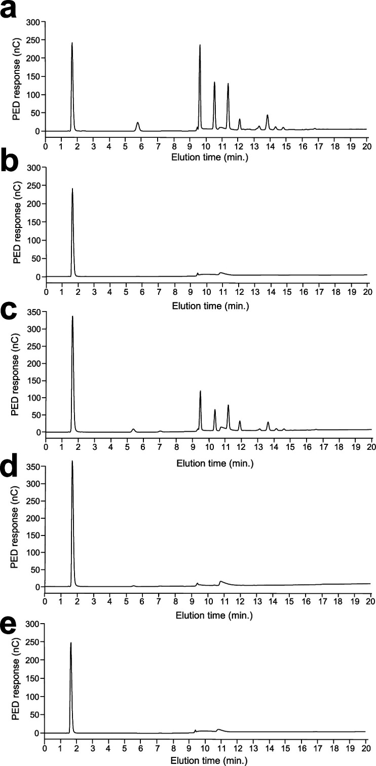
Figure 3—figure supplement 3.

High-performance anion exchange chromatography (HPAEC) analysis of oligosaccharides released by the endo-β-(1,6)-glucanase digestions of alkali-insoluble (AI) fraction of WT (a), AI fraction of kre6/kre62/skn2/skn1Δ/Δ (b), alkali-soluble (AS) fraction of WT (c), AS fraction of kre6/kre62/skn2/skn1Δ/Δ (d), and water (e). Experiments were performed in triplicates. PED, pulsed electrochemical detector; nC, nanocoulomb.