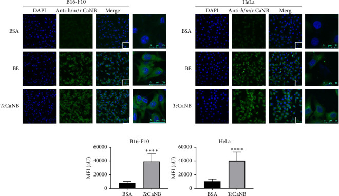
Figure 2.

Interaction of the recombinant TcCaNB protein with the cell surface proteins of B16-F10 and HeLa cells by an immunofluorescence assay. (a) B16-F10 and (b) HeLa cells were incubated with 5 μg/mL BSA (negative control), 10 μg/mL rat brain extract (BE), and 5 μg/mL TcCaNB for 1 h at 37°C. Cells were visualized by confocal microscope after incubation with polyclonal rabbit IgG Human/Mouse/Rat Calcineurin B antibody followed by goat anti-Rabbit IgG Alexa Fluor 488 (green) and DAPI (blue). Scale bar: 25 μm. (c) Quantification of the mean fluorescence intensity under BSA (negative control) and TcCaNB conditions. 100 cells were analyzed per condition. The results were expressed as average ± SD (Student's t-test, ∗∗∗∗p < 0.0001).
