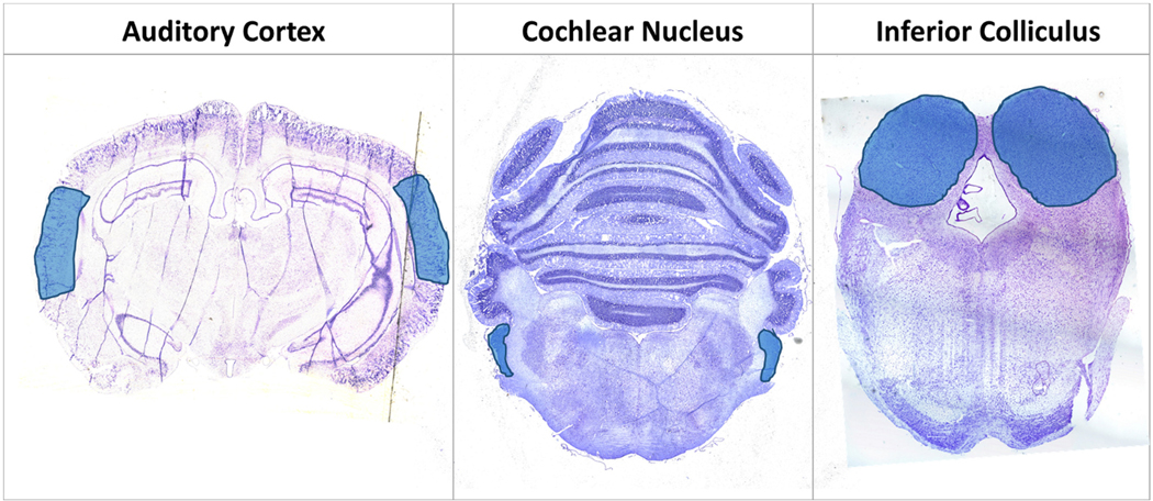
Fig. 3.
Cresyl-fast violet (CFV) stained 15 μm sections from the three areas of the primary central neuraxis within the sectioned slices of the male chinchilla brain. The blue areas are highlighting the auditory cortex (AC), cochlear nucleus (CN), and inferior colliculus (IC) regions of interest. (For interpretation of the references to colour in this figure legend, the reader is referred to the web version of this article.)

