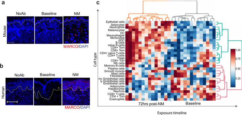
Fig. 6. Exposure of skin to nitrogen mustard results in the influx of MARCO-expressing cells.
Immunofluorescence staining of MARCO (red) in mouse (a) and human (b) skin sections. Mice were treated with nitrogen mustard and human subjects with Valchlor. Nuclei were stained with DAPI (blue). Dotted line indicates epidermal—dermal junction. Images were taken at 20× magnification, scale bar is equal to 100 µm. c shows the cell enrichment analysis of human skin biopsies taken at baseline and 72 h post-NM (Valchlor) treatment.

