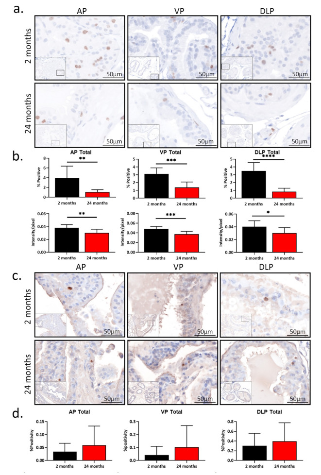
Fig. 4.
Total proliferation decreases with age with no change in apoptosis in mouse prostate (a) Pictures of prostatic Ki-67 staining from immunohistochemistry in young (2 months, n = 9) and aged (24 months, n = 9) mice. (b) The percentages of Ki-67-positive cells and the intensity of Ki-67 protein expression in young and aged anterior prostate (AP), ventral prostate (VP), and dorsal-lateral prostate (DLP). *p < 0.05, **p < 0.01, ***p < 0.001, ****p < 0.0001. (c) Pictures of TUNEL assay in young (2 months, n = 9) and aged (24 months, n = 8) prostates. (d) The percentages of apoptotic cells in young and aged AP, VP, and DLP.

