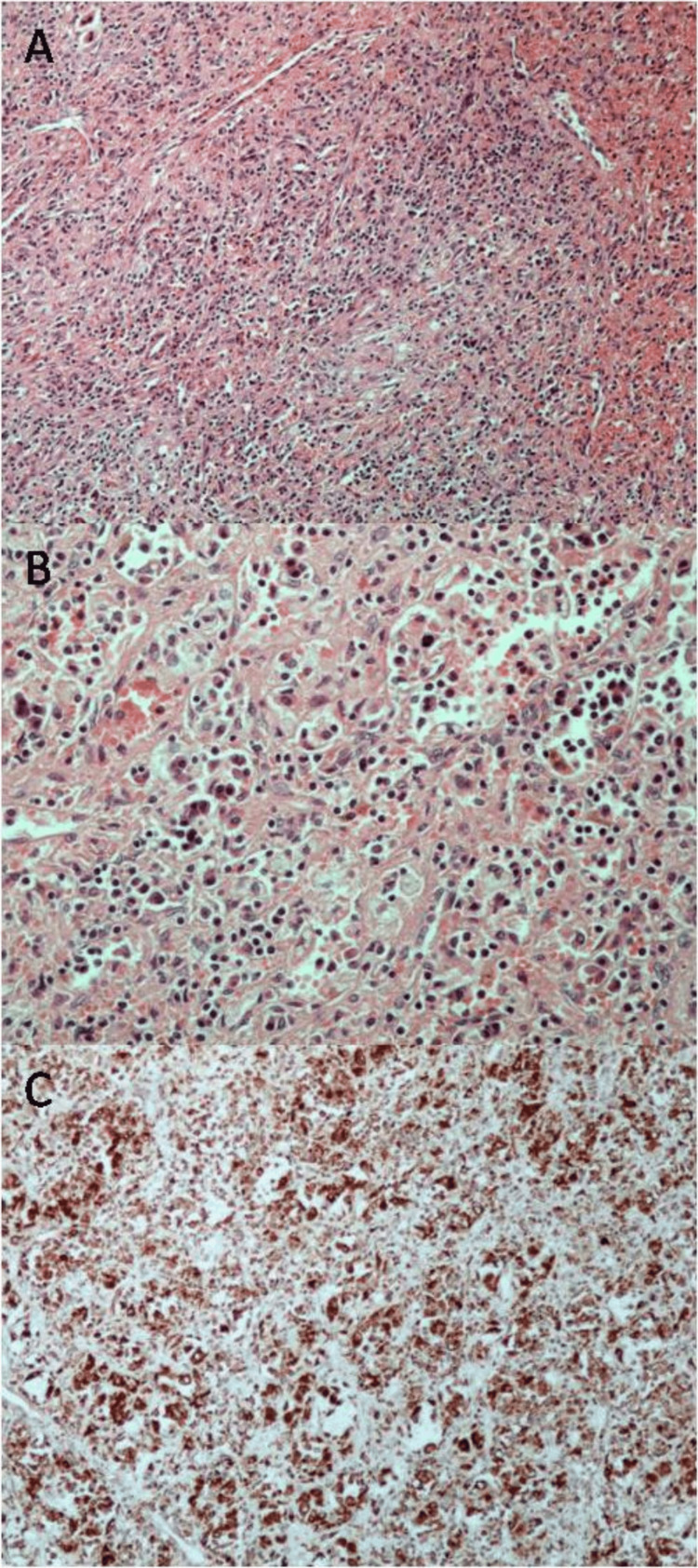
Figure 2. Histopathological features.
A, B. Red pulp substrate with fibroblastic reaction and moderate to severe inflammatory infiltrates, including "foamy" and iron-laden histiocytes, plasma cells, and lymphocytes; C. Immunohistochemical stain for CD68 highlighting histiocytes (A: hematoxylin-eosin, 100x; B: hematoxylin-eosin, 200x; C: streptavidin-biotin, 100x)

