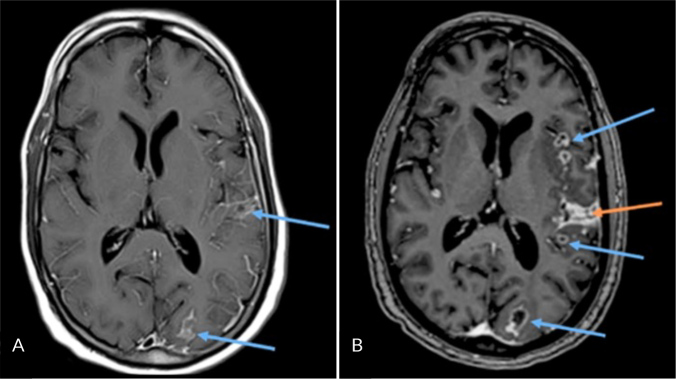
Figure 1.
A) Cerebral magnetic resonance imaging (cMRI) (T1 Blade after contrast administration) on the third day of hospitalization showing pachymeningeal enhancement (arrows) without amalgamation of cells; B) cMRI (T1-MPRAGE after contrast administration) three months after admission under continuing tuberculostatic therapy: progressive pachymeningeal enhancement (red arrow) and tuberculomas (blue arrow).

