Figure 2.

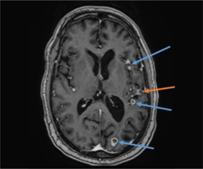
Cerebral magnetic resonance imaging (T1-MPRAGE after contrast medium administration) at 8 months after initial presentation. Size regression of the pachymeningeal enhancement (red) and the tuberculomas (blue).

Cerebral magnetic resonance imaging (T1-MPRAGE after contrast medium administration) at 8 months after initial presentation. Size regression of the pachymeningeal enhancement (red) and the tuberculomas (blue).