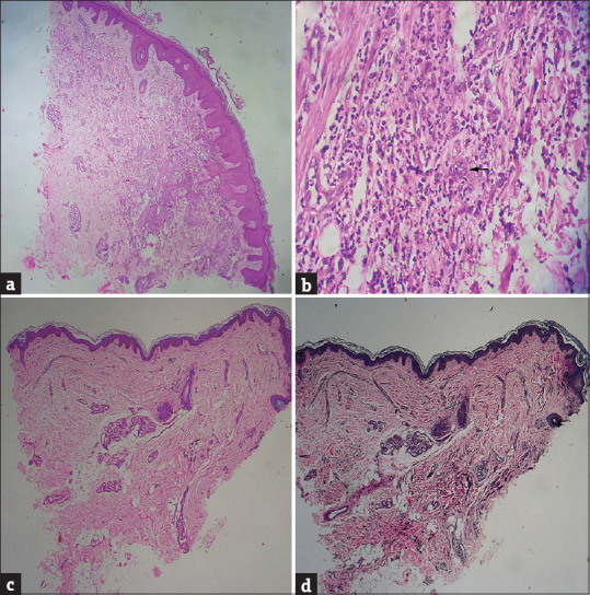
Figure 2.

(a) Diffuse dense infiltration of neutrophils in the upper dermis (H and E, ×40). (b) Significant leukocytoclasia along with dilated vessels and endothelial swelling (black arrow) (H and E, ×400). (c) Epidermal atrophy and absence of inflammatory cells in upper dermis after treatment (H and E, ×40). (d) Reduced elastin in upper dermis and coarse elastin fibres in lower dermis (VVG, ×40)
