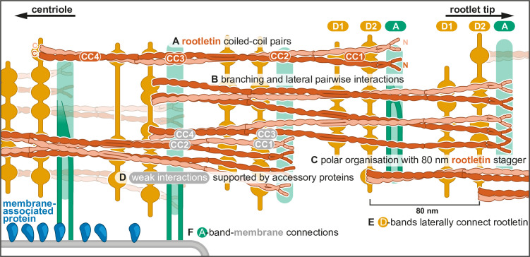
Figure 4. Model of rootlet organization.
(A) Filaments are depicted as rootletin coiled coils based on the 265-nm long AlphaFold prediction. Rootletin coiled coils are shown in different shades of orange for clarity. (B) Branchpoints are found where thicker filaments splay into thinner filaments, here indicated as single coiled coils. (C) Rootletin molecules are arranged in a polar manner based on the polarity we observed in the striations. The N-termini point away from the centriole, with an offset of 80 nm. We propose the N-termini align with the A-bands. (D) Staggered rootletin filaments may be supported via previously reported weak interactions supported by accessory proteins in the D-bands (E) D-bands were observed as punctate laterally connected densities associated with filaments. (F) Amorphous densities of the A-bands were occasionally observed to contain two parallel lines. A-band accumulations in purified rootlet suggest they correspond to the membrane interaction sites in cellular tomograms.

