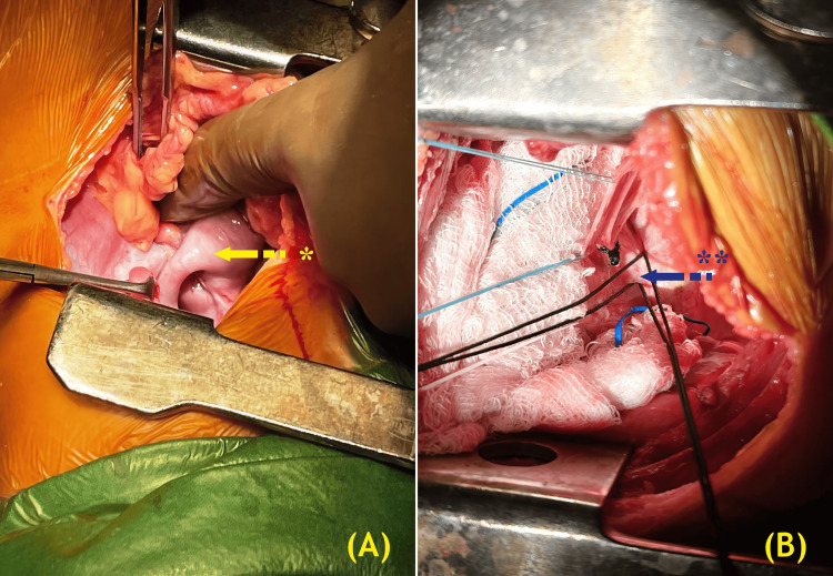
Figure 3. Right thoracotomy exposure showing (A) hernia contents, including the ileocecal appendix, cecum, ascending colon, and distal ileal loops. The yellow arrow and * indicate the appendix and small bowel in the right hemithorax. (B) PDA, looped before ligation during left thoracotomy exposure. The blue arrow and ** mark the PDA looped prior to ligation.
PDA, patent ductus arteriosus.
(Image credits: Dr. Vishal V. Bhende)

