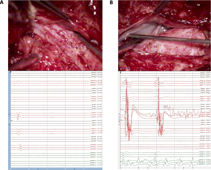
Fig. 4.
Intraoperative stimulation (upper row) and the corresponding intraoperative triggered EMG recording (lower row). Left column (A): After stimulation with the double-train paradigm, a single response in the TA, GAS, and abductor halluces muscles on the right side is recorded, indicating the sensory (posterior) S1 root (according to Schirmer et al. [34]). A small second response can be observed in the TA (and GAS), probably due to suprathreshold stimulation (4 mA) [27, 29]. Right column (B): After stimulation with the double-train paradigm, a double response in the adductor femoris, quadriceps, tibialis anterior (TA), and gastrocnemius (GAS) muscles can be recorded, with the highest amplitude response in the TA muscle on the right side indicating the motor (anterior) L5 root (according to Schirmer et al. [34])

