. 2024 Dec 4;11(1):e002394. doi: 10.1136/bmjresp-2024-002394

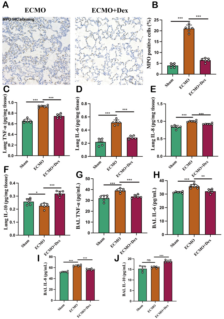
Figure 3. VV ECMO combined with Dex reduced pulmonary inflammation. Representative MPO immunohistochemical staining image (A) and comparison of MPO positive cells in the three subject groups (B). The level of TNF-α, IL-6, IL-8, and IL-10 in the lung (C–F). The level of TNF-α, IL-6, IL-8, and IL-10 in the BAL (G–J). The data shown are the mean±SD. Normality was analysed by Kolmogorov-Smirnov test. If the data were normally distributed, statistical significance was analysed by Student’s t-test, otherwise, a non-parametric Kolmogorov-Smirnov test was applied; ns=not significant, *p<0.05, **p<0.01, ***p<0.001. BAL, bronchoalveolar lavage; Dex, dexamethasone; VV ECMO, venovenous extracorporeal membrane oxygenation.

Figure 3