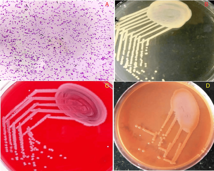
Figure 1. (A) Hematoxylin and eosin stain showing gram-negative, motile, polar flagellated bacterium suggestive of Stenotrophomonas maltophilia (magnification: 600 DPI; 400X). (B) Nutrient agar showing small circular, raised, yellowish pigmented colonies suggestive of Stenotrophomonas maltophilia (magnification: 600 DPI). (C) Blood agar classically showing non-hemolytic colonies of Stenotrophomonas maltophilia (magnification: 600 DPI). (D) MacConkey agar showing non-lactose-fermenting colonies of Stenotrophomonas maltophilia (magnification: 600 DPI).
DPI: dots per inch

