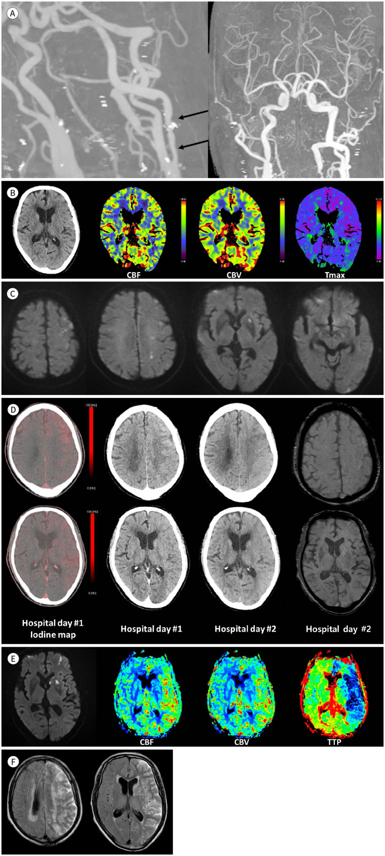
Fig. 1. An 80-year-old man with END as a form of CHS in acute ischemic stroke.
A. Brain CTA at the initial visit to the emergency department reveals moderate segmental stenosis in the left proximal internal carotid artery (arrows). Notably, there is patent flow to intracranial arteries with no evidence of occlusion or significant stenosis in intracranial arteries.
B. Brain CT and CTP at the initial visit to the emergency department. Non-enhanced brain CT shows no signs of intracranial hemorrhage and no clear hypodense area indicative of acute infarction. Brain CTP indicates symmetrical perfusion in both cerebral hemispheres, with no distinct perfusion abnormalities.
C. DWI during the initial visit to the emergency department. Multifocal tiny lesions with restricted diffusion, indicating acute infarction, are observed in the left frontoparietal cortex and white matter, left basal ganglia and left occipital cortex.
D. Dual-energy brain CT performed after trans-femoral cerebral angiography on the day of admission, follow-up brain CT and SWI on day 2 of hospitalization. The dual-energy CT obtained on the day of admission shows diffuse hyperdensities in the cortex of the left cerebral hemisphere, predominantly in the frontoparietal lobes. The follow-up brain CT, conducted on day 2 of hospitalization, reveals a reduction in the extent of diffuse hyperattenuation. In contrast, no hyperdense lesions are observed in the cortex of the right cerebral hemisphere, indicating that only the BBB in the left cerebral hemisphere is disrupted. Furthermore, the SWI performed on day 2 of hospitalization reveals no evidence of hemorrhage.
E. DWI and perfusion MRI on the 2nd day of hospitalization. DWI reveals a slight increase in the extent of acute infarction in the left cerebral hemisphere. Perfusion MRI demonstrates significantly increased CBF and CBV, along with decreased TTP in the left cerebral hemisphere compared to the right, indicating hyperperfusion.
F. FLAIR images on day 3 of hospitalization. FLAIR images show a high signal intensity along the cerebrospinal fluid space in the left cerebral hemisphere. This high signal intensity is presumed to be associated with the gadolinium contrast agent administered the previous day for the perfusion MRI.
BBB = blood-brain barrier, CBF = cerebral blood flow, CBV = cerebral blood volume, CHS = cerebral hyperperfusion syndrome, CTA = CT angiography, CTP = CT perfusion, DWI = diffusion weighted imaging, END = early neurological deterioration, FLAIR = fluid-attenuated inversion recovery, SWI = susceptibility weighted imaging, Tmax = time-to-maximum, TTP = time to peak

