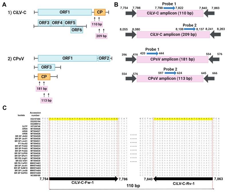
Fig. 1.
Designing of the primers and probes. (A) The coat protein coding region of 24 cytoplasmic-type CiLV-C isolates was aligned. Arrows indicate primer positions, and numbers indicate nucleotide positions on the consensus sequence. The size of the amplicon is shown below each alignment. (B) Genome organization of CiLV-C and CPsV. The CP region is boxed in orange, and arrows indicate the primer binding sites. (C) Detailed amplicons of each detection set. The binding sites of primers and probes are numbered based on the genomic location and orientation. CiLV-C, citrus leprosis virus; CP, capsid protein; CPsV, citrus psorosis virus; ORF, open reading frame.

