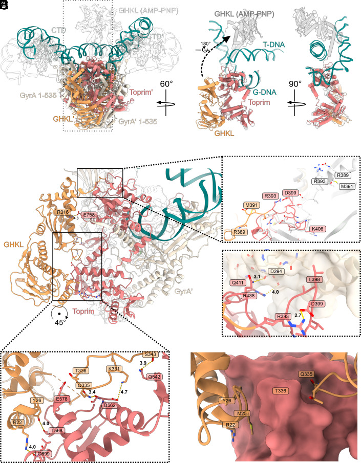
Fig. 2.
Position of the GHKL domain. (A) Superposition of Gyr-Mu217 (current work, PDB: 9GBV; color scheme as before) and E. coli gyrase in complex with 180 bp DNA, AMP-PNP, and gepotidacin [PDB: 6RKW (7); transparent contour]. Boxed region (a single GyrB subunit) is shown in isolation on the right to illustrate the extreme motion of the GHKL (12 nm shift and 180° rotation). AMP-PNP bound GHKL is shown in gray. (B) An overall view of the GHKL in the downward-folded conformation. Interactions with GyrA Tower and loop conformation (C and D) and interactions with the Toprim insert (E and F) are shown as insets. AMP-PNP-bound structure [PDB: 6RKW (7)] is shown as transparent contour or white cartoon [linker comparison between Gyr-Mu217 and PDB: 6RKW (7)].

