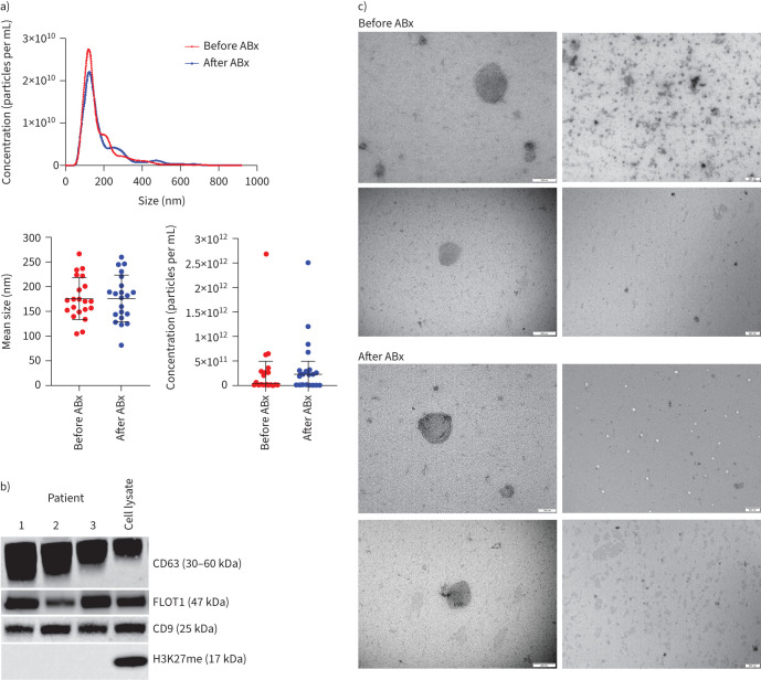
FIGURE 1.
Characterisation of extracellular vesicles (EVs) in sputum lysates: a) size distribution analysis of EVs before antibiotics (ABx) (red) and after ABx (blue) by nanoparticle tracking analysis (NTA); b) Western blot with anti-CD63, anti-CD9 and anti-FLOT1 antibodies, used as EV markers. CD63 core protein is a 26-kDa protein, the antibody detects the various glycosylated forms ranging from 30 to 60 kDa. Western blot with H3K27me (a tri-methylation of lysine 27 on histone H3 protein) was used as a control, to rule out cell debris in EV extracts; and c) transmission electron microscopy (TEM) for the morphologic characterisation of EVs in sputum homogenate. The two different magnifications highlight the morphology of individual EVs at narrow fields (left column, scale bars=100 nm (top) and 200 nm (bottom)) and a population of EVs at wide fields (right column, scale bar=500 nm).

