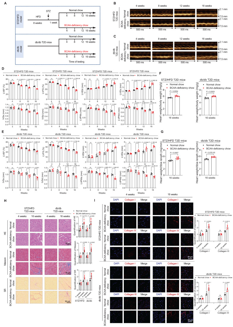
Figure 2.
BCAA deficiency time-dependently impairs cardiac function in STZ/HFD and db/db T2D mice. A, Schematic representation of the establishment of the two T2D models. B,C, M-mode echocardiogram images of STZ/HFD T2D mice (B) and db/db T2D mice (C) at four-week intervals after BCAA-deficient chow feeding (n=6 mice in each group). D,E, Echocardiography analysis illustrating the worsened heart function in STZ/HFD T2D mice (D) and db/db T2D mice (E) after BCAA-deficient chow feeding (n=6 mice in each group). F,G, Ratios of heart weight to body weight (F) and heart weight to tibia length (G) in indicated groups after 16 weeks of BCAA-deficient chow feeding (n=6 mice in each group). H, Hematoxylin and eosin, Masson's trichome, and Sirius Red stainings of heart tissues in indicated groups after 16 weeks of BCAA-deficient chow feeding. The image quantification is shown on the right (n = 5 mice in each group). I, Increased levels of Collagen I and III as detected via immunostaining of heart tissues from STZ/HFD and db/db T2D mice after 16 weeks of BCAA-deficient chow feeding. The image quantification is shown on the right (n = 5 mice per group). Data are expressed as mean±SEM. The nonparametric two-tailed Student's t-test was used to compare groups. Significance is indicated as nsP > 0.05, *P < 0.05, **P < 0.01, ***P < 0.001, and ****P < 0.0001. See also Figures S2 and S3.

