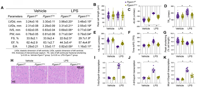
Figure 10.
Pgam1 deletion attenuated endotoxemia-mediated myocardial damage. Cardiomyocyte-specific Pgam1 knockout (Pgag5cko) mice and its control literature Pgam1f/f mice were injected with lipopolysaccharide (LPS) at 10 mg/kg for 48 hrs to induce an endotoxemia myocardial model. Single cardiomyocytes were isolated from Pgam1cko mice and Pgam1f/f mice on a Langendorff apparatus and the mechanical properties of cardiomyocytes were measured. A. Echocardiography was used to determine cardiac function. B-G. Mechanical properties were measured in 100-120 cardiomyocytes per group. H. Presentative pictures of HE staining in heart tissues after LPS exposure. I-K. RNA were isolated from heart tissues and the transcription of Il-6, Mcp1, and Tnfα were detected by qPCR. Data are shown as mean ± SEM. In each group, four animals or four independent cell isolations were used. Each experiment was conducted with three replicates and the dots in each panel represent the outcomes of these replicate experiments. *p<0.05.

