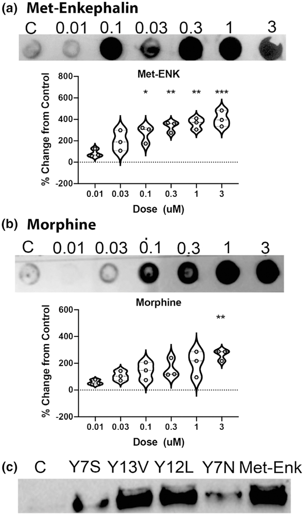
FIGURE 9.
esMOP MAPK signaling following agonist binding. (a) esMOP expressing HEK293T cells were serum starved overnight, and then treated with 0–3 μM Met-enkephalin or morphine for 5 min. Cell extracts were assayed by dot blot for phospho-MAPK, and intensity measured and expressed as percent change from control. Data represent means ± standard deviation, n = 3. (b) esMOP expressing HEK293T cells were cultured as above and treated with putative hagfish opioid peptides from esPENKL1: Y7S: YGGFMRS, Y13V: YGGFMRRFFGVAV, Y12L: YGGFMRRVGGPL, Y7N: YGRFMSN (1 μM) or Met-Enkephalin (Met-Enk; 1 μM) for 5 min. Phospho-MAPK was assessed with Western Blot

