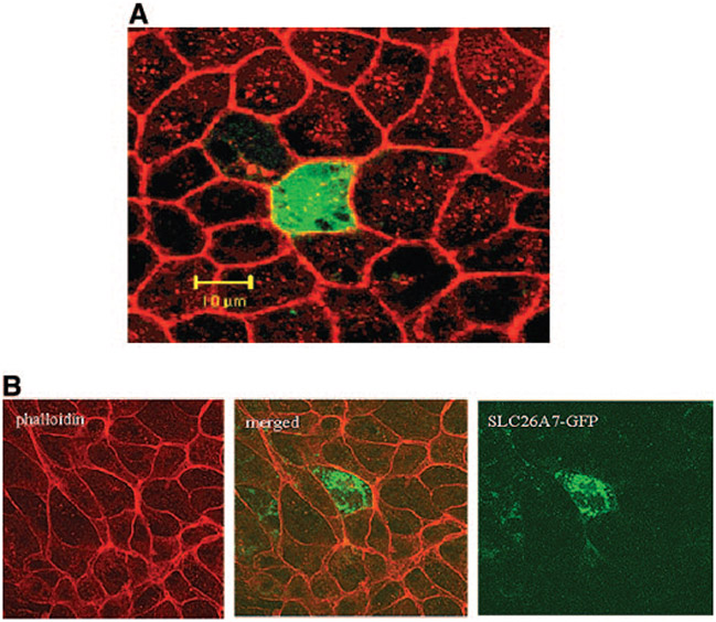
Figure 2.
Expression and subcellular distribution of green fluorescence protein (GFP)-SLC26A7 in MDCK cells. (A) Transfection with GFP vector alone (no SLC26A7 insert) results in GFP accumulation in the cytoplasm (Z-line images). (B) GFP-SLC26A7 is expressed in punctate cytoplasmic structures, with no labeling on the membrane. Red, phalloidin; green, GFP.

