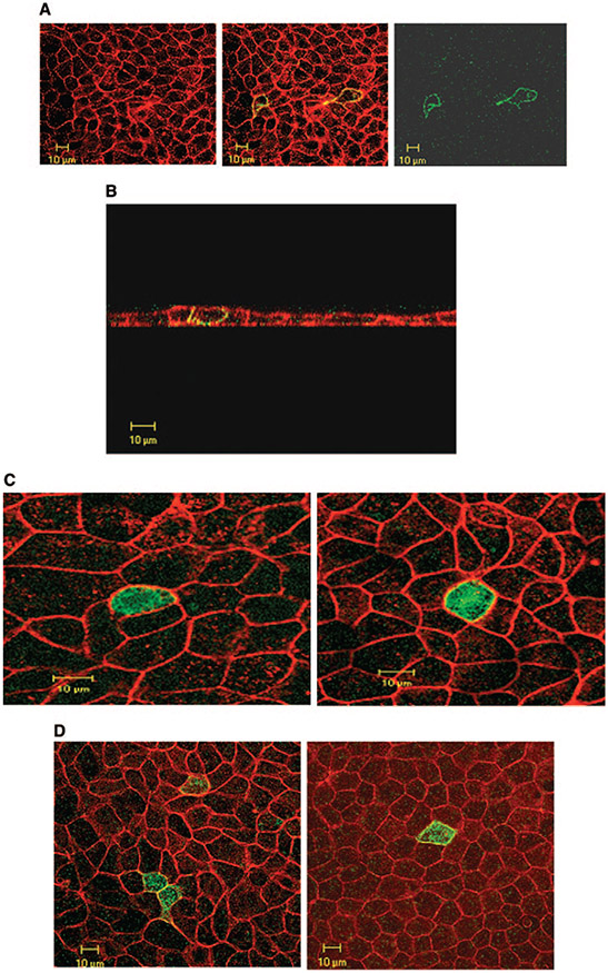
Figure 6.
Expression of SLC26A1, SLC26A7/A1 chimera, and truncated SLC26A7 in MDCK cells. (A) Expression of SLC26A1 in MDCK cells (Z-line image). SLC26A1 is expressed predominantly in plasma membrane, with no expression in the cytoplasm. (B) Subcellular distribution of SLC26A1 in isotonic medium (Z-stack images) indicates that SLC26A1 is localized to the basolateral membrane. Red, phalloidin; green, SLC26A1-GFP. (C) Expression of SLC26A7/A1 chimera in MDCK cells in isotonic and hypertonic media (Z-line image). SLC26A7/A1 chimera is expressed predominantly in the cytoplasm with some labeling in the plasma membrane (left, isotonic medium), and its distribution pattern does not change with increased osmolarity (right). (D) Truncated SLC26A7. As shown, the C-terminal–truncated SLC26A7 shows cytoplasmic distribution in isotonic medium (left). The truncated SLC26A7 remained predominantly in the cytoplasm with faint expression in the membrane in hypertonic medium (right).

