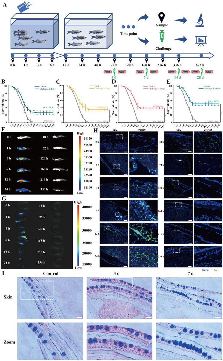
Fig 1. Strong immunoprotection in skin elicited by G3 against SVCV after immersion vaccination.
(A) Schematic diagram of in vivo uptake and metabolism analysis, sampling, and challenge of zebrafish after immersion vaccination by G3. Zebrafish were immersion vaccinated with G3 vaccine for 6 h. Subsequently, the vaccinated fish were transport to clean tanks. (B) After 3 days post-vaccination, the survival rates of immunized fish were tested for virus challenge. (C) After 7 days post-vaccination, the survival rates of immunized fish were tested for virus challenge. (D) After 14 days post-vaccination, the survival rates of immunized fish were tested for virus challenge. (E) After 28 days post-vaccination, the survival rates of immunized fish were tested for virus challenge. (F) Representative in vivo fluorescence images of vaccinated fish at different time points. The fluorescence intensity represents the amount of G3 vaccine detected. (G) Representative in vivo fluorescence images of skin in vaccinated fish at different time points. The fluorescence intensity represents the amount of G3 vaccine detected. (H) Representative in vivo fluorescence images of frozen section in skin at different time points. G3 vaccine were stained with FITC (green), nuclei were stained with DAPI (blue). Scale bars, 50 μm. (I) Histological examination by AB staining of skin from vaccinated fish at 3 dpv and 7 dpv. Scale bars, 50 μm. Fig 1A used some open-source artworks from Open Clipart and Wikimedia Commons. Science Beaker–Green (https://openclipart.org/detail/307196/science-beaker-green) and arrow next (https://openclipart.org/detail/12603/arrow-next) were used from Open Clipart under Creative Commons Zero 1.0 license (https://creativecommons.org/publicdomain/zero/1.0/), and no change were made to original artwork. Yellow test tube icon (https://commons.wikimedia.org/wiki/File:Yellow_test_tube_icon.svg), Microscope icon (black and blue) (https://commons.wikimedia.org/wiki/File:Microscope_icon_(black_and_blue).svg), and Analysis—The Noun Project (https://commons.wikimedia.org/wiki/File:Analysis_-_The_Noun_Project.svg) were used from Wikimedia Commons under Creative Commons CC0 1.0 Universal Public Domain Dedication license (https://creativecommons.org/publicdomain/zero/1.0/), and no change were made to original artwork. Syringe—Lorc—game-icons (https://commons.wikimedia.org/wiki/File:Syringe_-_Lorc_-_game-icons.svg) were was used from Wikimedia Commons under Creative Commons Attribution 3.0 Unported license (https://creativecommons.org/licenses/by/3.0/), and no change were made to original artwork. 201108 zebrafish (https://commons.wikimedia.org/wiki/File:201108_zebrafish.png) was used from Wikimedia Commons under Creative Commons Attribution 4.0 International license (https://creativecommons.org/licenses/by/4.0/), and no change were made to original artwork. Except for these open-source artworks, other parts in Fig 1A were drawn by ourselves.

