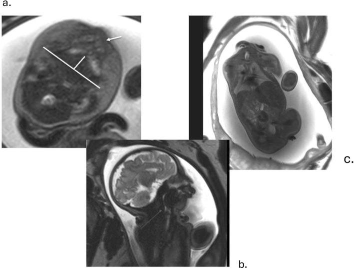
FIGURE 3.

Fetus at 36 weeks' gestation with micrognathia and cerebrocostomandibular syndrome due to an autosomal dominant de novo SNRPB pathogenic variant. (a) Axial T2‐weighted single‐shot EPI image demonstrating polyhydramnios and measurement of the mandibular depth, the numerator of the jaw index. Arrow indicates the alveolar ridge of the palate; note the posterior position of the mandible relative to the palate. (b) Sagittal T2‐weighted single‐shot EPI image demonstrating glossoptosis, resulting in marked narrowing of the oropharyngeal airway due to absence of the posterior palate consistent with Pierre Robin sequence. (c) Coronal T2‐weighted single‐shot EPI image showing bell‐shaped fetal chest due to rib deformities.
