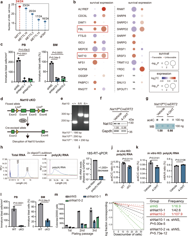
Extended Data Fig. 1. NAT10 is essential for AML.
(a) Number of AML cell lines with loss of fitness (CERES score lower than −0.5) when depleting specific genes. (b) Expression and survival analyses of TCGA-AML data showing the clinical relevance of the top 26 candidate genes with a CERES score lower than −1.0. (c) Histograms showing knockdown of Nat10 reduced the numbers and percentages of immature blast cells in peripheral blood (PB) and bone marrow (BM) from the primary BMT recipient mice. The number of immature blast cells were counted in 3 representative fields of Wright–Giemsa stained PB smear or BM cells. (d) The schematic diagram showing the strategy of conditional knockout of Nat10. (e) Genotyping of Nat10+/+, Nat10fl/fl or Nat10fl/+ mouse. (f,g) The reduction of Nat10 and RNA ac4C modification in the c-Kit+ BM cells of NAT10fl/flCreERT2 mice with or without in vitro 4-hydroxytamoxifen (4-OHT) treatment was confirmed by western blot (f) and dot blot (g) analyses, respectively. Cells were collected from the 1st passage of CFA. (h) Poly(A) RNA was isolated through two rounds of oligo(dT) pulldown and the purity was verified through size distribution in Qsep Bio-Fragment Bioanalyzer profiling. (i) Validation of poly(A) RNA purity through RT–qPCR with primers specific to 18S rRNA and GAPDH. (j) LC–MS/MS showing the reduction of ac4C modification levels in the poly(A) RNA of c-Kit+ BM cells from NAT10fl/flCreERT2 mice with in vivo tamoxifen treatment. (k) LC–MS/MS showing the ac4C modification levels (left) and m6A modification levels (right) in the poly(A) RNA of c-Kit+ BM cells from NAT10fl/flCreERT2 mice with in vitro 4-hydroxytamoxifen (4-OHT) treatment. (l) Histograms showing knockout of Nat10 reduced the numbers and percentages of immature blast cells in peripheral blood (PB) and bone marrow (BM) from the primary BMT recipient mice. The number of immature blast cells were counted in 3 representative fields of Wright–Giemsa stained PB smear or BM cells. (m) Colony numbers of mouse HSPCs transduced with MLL-AF9 plus shRNAs targeting Nat10 or non-specific control (shNS) in methylcellulose medium. (n) In vitro LDA in mouse HSPCs co-transduced with MLL-AF9 and shNS or shRNAs targeting Nat10. Values are mean ± s.d. of n = 3 biological replicates in c, i, j, k and l. Two-tailed Student’s t-tests were used. Values are mean of n = 2 biological replicates in m. Two-sided Chi-squared tests were used in n. Images in f and g were representative of three independent experiments.

